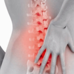
Signs of a herniated spine
Intervertebral hernia is a fairly common disease of the musculoskeletal system that occurs as a result of rupture of the fibrous ring of the intervertebral disc and displacement of its nucleus in the anteroposterior direction , which leads to the development of a typical clinical picture and symptoms of the disease.
Spinal hernia is a serious pathological condition that requires mandatory timely correction, otherwise this disease can lead to serious complications associated with pinching and atrophy of the nerve fibers of the spinal roots. In order to prevent the progression of the pathological condition and the development of complications, it is necessary to know the typical clinical signs and how a hernia manifests itself.
Medical tactics
In order to determine the presence or absence of a disease, it is necessary to know the signs of a spinal hernia. To do this, the specialist who treats the patient collects data on the anamnesis of life and the present disease, finds out the patient's complaints and determines the symptoms, after which he forms a preliminary diagnosis, which makes it possible to more accurately prescribe additional instrumental diagnostic studies. Only a competently conducted questioning and determination of pathological symptoms and syndromes allow us to understand the cause of complaints, as well as quickly and correctly establish a diagnosis.
Anamnesis
Already at the stage of collecting anamnestic data, the attending physician may suspect an intervertebral hernia. So quite often the disease is provoked by excessive physical loads on the musculoskeletal system, in particular, axial loads on the spinal column. There is a clear correlation between the occurrence of an intervertebral hernia and work associated with difficult working conditions. This disease is much more common in men of middle and older age groups, employed in production, road or loading operations. Even a long-standing injury to the vertebrae can provoke the development of protrusion and hernia in the future.
Family history data also help in the diagnosis, since in some cases there is a hereditary predisposition to degenerative diseases of the connective tissue, and when an intervertebral hernia occurs, a violation occurs in the composition of the connective tissue of the body.
Complaints
Patients suffering from a herniated disc can either show active complaints or say that nothing bothers them at all.
In most cases, patients complain of pain of varying intensity of aching nature approximately in the part of the spinal column where the hernial protrusion is located. In some cases, the patient does not associate complaints with pain in the spine at all, which may lead to incorrect treatment, as a result of a false diagnosis.
Quite often, patients resort to self-treatment. For a long time, they do not seek advice and qualified medical care, which leads to blurring of the clinical picture and the development of complications, such as radicular syndrome.
In addition to severe pain, patients may experience parasthesia - a feeling of goosebumps on the skin and a decrease in tactile sensitivity on the distal parts of the upper and lower extremities. In the most severe cases, the passage of electrical impulses along the fibers of the spinal roots is so difficult that the patient begins to experience constant muscle weakness - paresis. Signs of a spinal hernia can develop over a long period of time, in some people a hernia may appear years after the rupture of the annulus of the intervertebral disc.
Symptoms
Let's take a closer look at how an intervertebral hernia manifests itself in order to suspect a disease already in the early stages of formation. Depending on the location of the hernia in the human spinal column, the symptoms of this pathological condition will change.
Intervertebral hernia can be in three anatomical regions of the spinal column:
- In the cervical region . Such hernias are quite rare due to the anatomical features of the structure of the cervical vertebrae and ligamentous apparatus, as well as due to the absence of high loads on this section of the spinal column. However, under certain circumstances, an intervertebral hernia can still form in the cervical region.
- In the chest. Intervertebral hernia of the thoracic region occupies a middle position in the structure of morbidity between hernias of the cervical and lumbosacral regions. The load on this section of the spinal column is higher than on the cervical one, however, a feature of its structure is rigid fixation due to the formation of the chest.
- In the lumbosacral region. The most common type of intervertebral hernia, since this department accounts for almost the entire volume of axial load. It is in the lumbar intervertebral discs that dystrophic processes most often develop, eventually leading to a displacement of the central nucleus of the intervertebral disc.
radicular syndrome
One of the most common complications of a herniated disc, associated with prolonged compression of the nerve fibers leaving or entering the spinal canal. The radicular syndrome is manifested by a pronounced pain syndrome with different localization of pain sensations.
Prolonged compression of the nerve roots in the projection of the hernia leads to their inflammation, which leads to the development of local edema, thereby closing the vicious circle of radicular syndrome. Ultimately, the radicular syndrome, which has developed as a result of a herniated protrusion of the intervertebral disc, leads to the development of muscle atrophy of the innervated limbs and impaired movements in them, since the spinal roots include motor fibers of the spinal cord motor neurons responsible for contraction of striated muscle tissue and our movements. Radicular syndrome localized in the cervical spine leads to pain along the inflamed nerve, thus, patients complain of pain in the neck and arm on the side of the affected root. During the formation of radicular syndrome in the thoracic region, pain can be visceral in nature and appear in the chest cavity in the form of cardialgia and gastralgia, which complicates preliminary diagnosis. If the syndrome develops in the lumbar or sacral region, then often patients are disturbed by severe acute pains resembling backaches. Pain is given to the lower limb on the side of the affected spinal root. In some cases, the pain is so strong that the patient is forced to take a certain position to reduce the intensity of pain.
How to determine a hernia of the spine, depending on the location?
Cervical hernia
A vertebral hernia that has formed in the cervical region can be manifested by the formation of symptoms such as:
- Frequent headaches resulting from impaired blood flow through the vertebral arteries and feeding a large part of the cerebellum;
- Dizziness not associated with disorders of the vestibular apparatus;
- Increased intracranial pressure due to a violation of the drainage function of the cerebrospinal fluid in the spinal canal;
- Noise in ears;
- Numbness predominantly of the upper extremities.
If you managed to identify similar symptoms, then you should not postpone visiting a specialist traumatologist-orthopedist, since only professional medical advice and differential diagnostics can answer the question of whether the patient has a hernia or not.
Thoracic hernia
For a hernia of the thoracic region in the symptoms, other symptoms come to the fore:
- Pain syndrome. The severity of pain increases gradually, in proportion to the progression of the displacement of the intervertebral nucleus. The pain is localized in the thoracic region, while most often the patient fails to accurately point to the sore spot, so the pain is widespread throughout the thoracic region.
An important point in the formation of pain as a result of an intervertebral hernia in the thoracic region is the masking of symptoms under other diseases, such as coronary heart disease or stomach ulcers. This is due to the peculiarity of the innervation and exit of sensitive nerve fibers from the spinal roots located in the thoracic spine.
It should be noted that in some cases, when the spinal column is injured, the symptoms may be inactive for a long time, which is associated with the compensatory mechanisms of the body. In such cases, the victim can live up to several years without even realizing such a serious disease as a herniated disc.
Hernia of the lumbosacral region
This type of hernia is the most common and is associated with a high degree of stress on the lumbar and sacral regions along the vertical axis. The hernia is formed slowly as a result of degenerative changes in the connective and bone tissue of the lumbar region, which leads to thinning of the fibrous ring and displacement of the intervertebral nucleus. In most cases, patients develop pain syndrome of varying intensity. Pain sensations gradually increase and are localized at the beginning of the disease in the lumbar region, and then begin to radiate to the buttocks and lower limbs.
Diagnostics
How to determine the intervertebral hernia? Diagnosis of intervertebral hernia is a mandatory step before starting treatment, since only a correctly established clinical diagnosis can help the attending physician find out and form the correct and effective treatment.
In addition to the symptoms described above, it is necessary to conduct a complete differential diagnostic study, not only to establish a clinical diagnosis, but also to establish the exact localization of the pathological process. A spinal hernia may not make itself felt for a long time and is detected in patients as a diagnostic finding, for example, during an X-ray examination or MRI for some other disease.
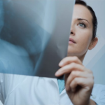
What does a hernia look like on diagnostic examination? For a detailed diagnosis of an intervertebral hernia, magnetic resonance imaging, or MRI for short, is used. MRI helps to understand exactly where the hernial protrusion is located and how much it has a compressive effect on the nerve tissue of the spinal cord. Unlike radiological research methods such as plain radiography and computed tomography, magnetic resonance imaging allows you to show in detail the condition of soft tissues, which is valuable information for determining the degree of damage to the spinal roots subjected to compression from the hernia.
What does a herniated disc look like on an MRI? When conducting a tomographic study on the patient's image, specific changes characteristic of a hernia are revealed. These signs include:
- Stretching or rupture of the fibrous ring of the intervertebral disc on an MRI image. If there is no rupture, then this condition is called protrusion or prolapse.
- As a result of flattening of the intervertebral disc on the tomogram, the displacement of the nucleus pulposus of the intervertebral disc becomes visible towards the spinal canal, or into the lateral projection.
- Compression of the roots and inflammatory tissue edema allows you to find out if there is a radicular syndrome in each case.