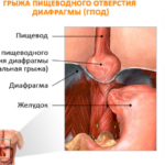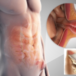
What is a diaphragmatic hernia
Diaphragmatic hernia is a disease that occurs as a result of the movement of a certain part of the esophagus, stomach or other elements of the gastrointestinal tract into the chest cavity through a hole in the diaphragm. The incidence of such a pathology among all hernias is on average 2%. Most often, such a disease is detected when people complain of pain and other disorders in the gastrointestinal tract.
The diaphragm is the main muscle involved in the breathing process. On the other hand, this muscle plays the role of the abdominal septum, delimiting the abdominal cavity from the chest. This anatomical formation is located directly under the lungs and is attached to the ribs.
The muscular organ consists of two parts - the central tendon component and the muscular peripheral element. It is in the second that the natural opening for the esophagus is located. It is the weak point through which, most often, diaphragmatic hernias are formed.
With a significant increase in internal pressure, the organs located in the abdominal cavity (stomach, sometimes intestines) and the esophagus leave their anatomical location and pass through the hole in the respiratory muscle, ending up in the chest cavity.
Causes of a hernia
The main factors in the formation of the disease include:
- genetic defects in the development of the diaphragm in newborns;
- prolonged increase in pressure in the peritoneum against the background of chronic cough, strength exercises, heavy physical exertion, gestation, constipation, obesity;
- intravital injuries : penetrating wounds of the peritoneum, blows, falls on the stomach;
- age - most often, diaphragmatic hernias occur in people over 50 years old, which is associated with physiological changes in the muscle;
- transferred diseases of the nervous system , in which the phrenic nerve was damaged, which led to a violation of the innervation of the muscle;
- background diseases of the gastrointestinal tract : inflammation of the esophagus, cholecystitis, peptic ulcer of the stomach and intestines, diseases of the pancreas.
Classification of diaphragmatic hernias
There are several types of diaphragmatic hernia.
They are divided into four groups:
- Congenital hernia . It is formed more often than others, and is associated with genetic anomalies in the development of the baby.
- neuropathic . This type occurs against the background of impaired muscle tone. In this case, the diaphragm relaxes, and this contributes to the stretching of the muscle fibers, which can lead to rupture and the formation of a bulge.
- Traumatic hernia . It happens to both children and adults. This type of pathology exists in two versions, namely: true and false hernia. It is formed due to any injury to the respiratory muscle.
- A hernia formed by a natural opening in a muscle . In the case of stretching of tissues of any nature, the natural opening can expand in diameter, which makes it possible for the organs to enter the chest cavity.
True diaphragmatic hernia
With this variant of the pathology, there is a hernial sac. Its wall is thin, devoid of any muscle fibers. A favorite place for the formation of such hernias is "weak" spaces, otherwise - Larrey's gaps. One of the variants of this disease are hernias of atypical localization, that is, those places where the location of the hernia is not typical for the standard case. This kind is extremely rare.
False diaphragmatic hernia
The congenital variant of the false protrusion is considered a malformation. A hernia in this case is formed due to a violation of the fusion in the embryonic period of the connections between the abdominal and chest cavities. The absence of a hernial sac is a hallmark of the false variant of the pathology, while there is a through hole in the respiratory muscle.
Diaphragmatic hernia symptoms
The characteristics of the symptoms depend on the nature and type of hernia.
Also, the signs of the disease are determined by the following factors:
- rate of disease development : acute or chronic;
- the duration of the hernia in an abnormal position;
- the presence of complications (pinching, inflammation).
The clinical picture of acute hernias:
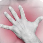
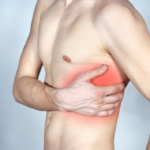
- Constant pain in the chest , resulting from mechanical compression of the organs. Pain is aggravated by coughing.
- Heartburn , aggravated by the horizontal position of the body. Also, the feeling of heat increases when the body bends down. In addition, heartburn can occur after eating.
- Belching , it can be in two versions, namely: belching with air or sour. This phenomenon is also present during sleep.
- Difficulties in the act of swallowing . Swallowing, the patient every time feels a lump in the chest region. This symptom is associated with the intake of both liquid and solid food.
- Flatulence and bloating.
- Chronic dry cough.
- Heaviness when breathing . The patient may complain of shortness of breath, as if he does not have enough air, or he cannot breathe.
- Sensation of strong palpitation after eating.
- Unusual sounds such as gurgling in the chest area.
The chronic variant of diaphragmatic hernia suggests a latent nature of the development of symptoms, that is, at first the patient does not feel a painful condition. The height of the symptoms appears later, and the clinical picture corresponds to an acute course. The course of diaphragmatic hernia in children does not differ from that in adults.
Establishing diagnosis
Diagnosis of the disease is based on data from four main studies.
Diagnostic methods:
- X-ray examination of the cavity of the chest and peritoneum . In real time, X-rays allow you to see the dynamics of the movement of barium through the alimentary tract.
- Fibrogastroscopy . The study is achieved by introducing a thin tube with a camera at the end into the stomach, while passing through the esophagus itself. Thanks to FGS, doctors can see various complications such as bleeding.
- pH measurement.
- Intravital tissue sampling - biopsy . However, this method is prescribed only when absolutely necessary.
Diaphragmatic hernia treatment
Diaphragmatic hernia can be completely cured only by surgical intervention. However, this pathology is famous for its relapses: in about half of the cases, the protrusion reappears, so the priority treatment is conservative therapy.
This type of therapy is aimed at preventing and inhibiting the development of the inflammatory process, and the tasks include: preventing disorders of the gastrointestinal tract and normalizing pressure in the abdominal cavity. In addition, background diseases such as gastritis or ulcers are cured in parallel.
There are two main methods of conservative treatment:
- Diet and its rules:
- exclusion from the diet of simple carbohydrates that can cause fermentation in the intestines;
- the exclusion of acidic foods, such as juices, citrus fruits, berries - all that can provoke damage to the gastric mucosa;
- the exclusion of products that initiate abundant secretion of hydrochloric acid or pancreatic enzymes. These foods include: fried and smoked foods, spicy flavors, pickled vegetables;
- inclusion in the daily diet of dried fruits. A special place among them is occupied by prunes;
- the use of alkaline mineral waters;
- do not take a horizontal position after eating and do not go to bed.
- The second method is drug treatment. Basic goals:
- reduction of excessive production of gastric juice. Achieved by taking "atropine" or "platifilin";
- elimination of hypertonicity of the abdominal muscles, which leads to a weakening of the pain syndrome. Antispasmodics are used;
- prevention of the destructive effect of gastric acid on the mucous membrane. For this, enveloping agents such as "De-Nola" are prescribed.
Another way to treat diaphragmatic hernia is surgery. It is prescribed in 10% of all cases of the disease.
The team of surgeons has two tasks:
- Elimination of hernial ring.
- Creation of a barrier that prevents the sudden throwing of hydrochloric acid into the lumen of the esophagus.
The operation consists in the displacement of the esophagus and stomach from the chest cavity towards its anatomical position. This goal is achieved by two methods: laparotomy (entrance through the peritoneal cavity) and thoracotomy (entrance from the side of the chest).