Signs and treatment of paramedian disc herniation L4-L5
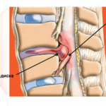
Paramedian l4 l5 disc herniation is a pathological condition that manifests itself as a degenerative lesion of the cartilage of the intervertebral discs with their partial or complete displacement into the spinal canal, which subsequently leads to compression of the latter in the lumbar region. The danger of the disease is when the protrusion exerts compression not only on the spinal cord, but also on the outgoing nerve roots.
Often, the disease is formed against the background of the course of degenerative diseases of the musculoskeletal system (congenital dysplasia, osteoarthritis or osteoporosis).
Why does a paramedian hernia appear?
Lumbar hernia is quite common. This occurrence is explained by the fact that the lumbar region takes the greatest load of the whole body on itself. Under prolonged exposure to strong pressure, the core of the intervertebral disc undergoes a number of organic changes, namely: the core “dries out” and eventually collapses, thereby losing its former elasticity.
In the future, the central part can no longer absorb the applied pressure, and the peripheral part of the disk goes into an area of less resistance.
The main causes of the development of the disease:
- a lifestyle associated with strong physical exertion on the back of a person;
- excess body weight;
- transferred operations on the spine;
- back and muscle injuries;
- hereditary predisposition, which determines the weakness of one or another part of the back (muscles, ligaments);
- age-related changes in the human skeletal system;
- ankylosing spondylitis;
- scoliosis, lordosis or kyphosis;
- diseases of the endocrine system leading to an imbalance of minerals in the human body;
Varieties
There are several types of hernias. These are classified according to different criteria.
Depending on the location relative to the midline of the spine, there are:
- Lateral (located on the sides - left or right) - when the pathological process involves the lateral parts of the intervertebral disc. Right -sided paramedian hernia is formed not only in the lumbar, but also in the cervical region. L e lateral is often directed inward the spine.
- Dorsal (posterior) - the back of the fibrous disc is affected, followed by bulging into the paramedial space of the spine.
- Sequestered - the hernia completely falls into the lumen of the canal of the spinal column with gross compression of the spinal cord and nerve roots.
Clinical picture
The peculiarity of the symptoms is determined by the anatomical structure of the spinal column and spinal cord. The leading symptom of the disease is pain, which is associated with squeezing of the bulge on the nerve endings coming out of the spinal cord. The pain has a versatile character: it can spread not only to the back, but also to the lower limbs, give to the pelvic area. Pain sensations are sharp in intensity, shooting through.
It should be remembered that the pain can be aggravated by coughing, sneezing, bending over.
Signs of a left-sided lateral hernia:
- Phenomena of dysfunction of the working capacity of the arm or leg on the left side. Also in these places, the patient may feel numbness and decreased sensitivity. The work of reflexes is disrupted: they can decrease or disappear completely.
- General cerebral symptoms : vomiting, dizziness, severe headaches, periodic nausea. The patient feels a deterioration in well-being, his performance decreases.
Right-sided hernia has the same symptoms, but most of them appear on the right side of the body. Patients complain of disruption of the pelvic organs. Painful and difficult urination is observed, libido decreases in men, erection function worsens. In severe cases, spontaneous urination may occur.
A volumetric hernia can strongly compress the outgoing roots, which leads to compression, edema, and blood stasis. In extreme cases, aseptic inflammation may occur.
Diagnostic measures
To make the correct diagnosis, the doctor resorts to various methods of examining the patient. He studies the signs of a violation of the nervous activity of the body: motor, sensory, reflex and autonomic disorders. To do this, the specialist conducts a general examination, studies the anamnesis of life and illness. A direct study of the pathology is carried out with an objective study, then the doctor, using neurological instruments, studies the spectra of dysfunction.
Another stage in the diagnosis is the use of instrumental methods.
These include:
- Radiography . Here, the attending physician will be able to see various changes in the configuration of the spine, an individual vertebra, their shifts, deformation, flattening, narrowing of the spinal canal or thickening of the connective tissue.
- Magnetic resonance imaging . This method gives visualized information about how the disc or its elements are damaged, information about the degree of compression of the roots.
- Myelography . This method informs the doctor about the ability of the muscles to contract, about their innervation. To
- Computed tomography studies the parameters of a hernia: its size, displacement, contact with neighboring tissues or structures, and the direction of the hernia nucleus. The method itself is based on the use of x-rays.
Treatment
Any disease is treated in two ways: conservative treatment and surgical intervention. After the diagnosis and diagnosis, conservative therapy is a priority.
Her principles:
- Treatment without intervention is most effective when it begins in the early stages of the disease. The main means at this stage are anti-inflammatory, decongestant and analgesic drugs.
- The hospital may use drugs containing L-lysine aescinate or eufillin (if the patient's body has no contraindications). Also spazmaton or tolperizol. These drugs are aimed at relieving muscle spasm, which leads to their relaxation and "release" of nerve endings. This helps relieve pain.
- Anti-inflammatory drugs are aimed at removing inflammatory processes and edema , prevent the destruction of nerve roots, and partially relieve pain. If there are signs of severe edema, the doctor prescribes diuretics, and with severe pain, drugs containing hormone elements.
- With a stable course of pathology, therapeutic exercises, massage and manual therapy are indicated. These are all muscle relaxation and recovery techniques that reduce the damaging effect on pinched nerves.
- Physiotherapeutic procedures : ultrasonic wave therapy, phonophoresis.
Surgical treatment is used in cases where conservative therapy had no effect, or the patient is in serious condition. Also, surgery is a priority when the hernia is large, reduces the spinal canal.
Invasive treatment pursues several tasks: to release the nerve roots from compression, to eliminate pathological tissues, that is, the hernia itself.
A common method is discectomy. Treatment with this method involves the creation of several small incisions through which the separation of protruding sections of the intervertebral disc is carried out and their further removal using an endoscope. The advantage of such an operation is that the patient who underwent the intervention will be able to walk the very next day.
Postoperative period and rehabilitation
In the first time after therapeutic measures, the patient requires rest, deprivation of physical activity. It is recommended to wear a medical bandage in the form of a corset. This period is up to a month. In the future, a cured person needs to dose the amount of physical activity, eat right (nutrition involves the inclusion of vitamin complexes in the diet), continue therapeutic exercises.
Disease prevention
Proper and rational exercise of dosed physical culture occupies a special place in the prevention of not only hernias, but also degenerative diseases of the musculoskeletal system. It is necessary to monitor weight, control sudden changes, engage in exercises that are aimed at developing the mobility of the spinal column and strengthening the ligamentous system.


